
Our Technology
iTero Scanner
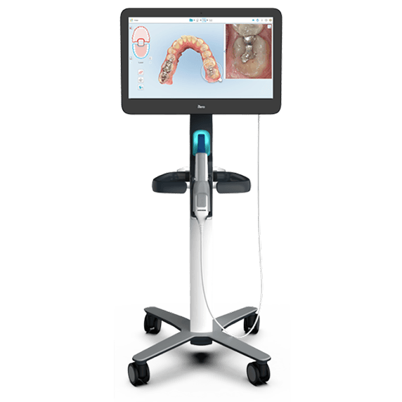
Remember the putty-filled trays that made you gag while you counted the seconds? We don’t use those. The iTero scanner is a small handheld wand that takes thousands of images of your teeth and gums and stitches them into a detailed 3D model in a matter of minutes. Nothing sets, nothing hardens, and you can swallow and breathe normally the whole time.
That digital model does a lot of work behind the scenes. It’s what your Invisalign aligners are built from, and because the scan is accurate down to fine detail, your aligners and appliances fit the way they’re supposed to from the first tray. If something needs a second look, we rescan on the spot instead of mailing a physical mold across the country and waiting.
The part patients tend to like most is seeing it. Your scan appears on the screen right next to you, so our doctors can rotate the model, point out the crowding or the bite issue they’re describing, and walk you through the options — braces or Invisalign — with your own teeth as the visual aid. We keep your scans on file, which makes it easy to compare where you started with where you are partway through treatment.
A lot of orthodontics happens where you can’t see it — roots, developing teeth, and the way your jaws are positioned relative to each other. Panoramic and cephalometric X-rays give our doctors that view, and they ask very little of you to do it. Both are digital, so each image takes a small fraction of the radiation film once required — a panoramic dose is roughly what you’d absorb from the environment over a day or two. Nothing goes in your mouth, the whole thing takes seconds, and the images are up on screen immediately rather than at a follow-up visit.
A panoramic X-ray (the “pan”) captures your entire mouth in one wide image: all of your teeth, both jaws, your sinuses, and your jaw joints. It shows teeth that haven’t come in yet, teeth growing in at the wrong angle, extra or missing teeth, and how much room there is for everything to fit. For kids and teens, this is often what tells us whether to start treatment now or wait a year.
A cephalometric X-ray (the “ceph”) is a side-profile view of your head. It shows how your upper and lower jaws relate to one another, how your teeth are angled, and how your face is growing. Our doctors use it to decide whether a bite problem is coming from the teeth, the jaws, or both — which is the difference between a straightforward treatment plan and one that needs a different approach entirely.
Our doctors take these images only when the result will actually inform a treatment decision, never as a routine box to tick. They’ll pull them up and go through them with you as part of your free consult, so you can see what they’re seeing and understand the plan before treatment starts.